Pulmonary Disease Pattern On Ekg
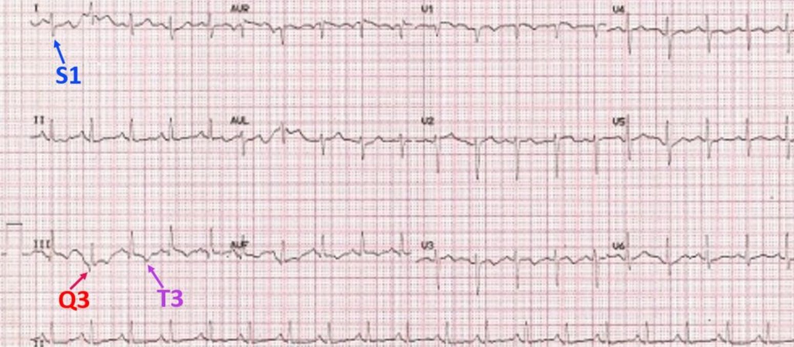
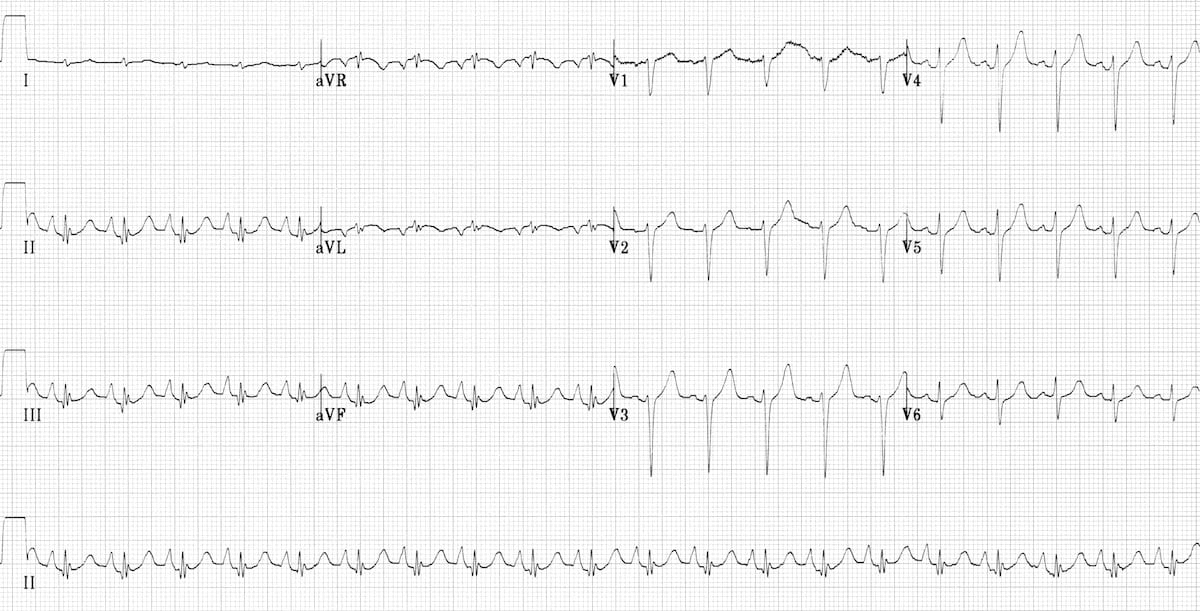
Pulmonary Disease Pattern On Ekg - Web chronic obstructive pulmonary diseases (copd), a broad spectrum of respiratory diseases represents a worldwide problem. Web ecg abnormalities are common in patients with pulmonary embolism, with the most frequent being sinus tachycardia, right ventricular strain, and the classic s1q3t3 pattern. Web aggregation of data from echocardiography, heart catheterisation and spirometry allowed us to relate ecg patterns in copd to the separated, graded effects of emphysema, airway obstruction and rv afterload. Ecg findings often suggest right ventricular pressure overload or strain. Web electrocardiography (ecg) is a useful adjunct to other pulmonary tests because it provides information about the right side of the heart and therefore pulmonary disorders such as chronic pulmonary hypertension and pulmonary embolism. Increased stimulation of the sympathetic nervous system due to pain, anxiety and hypoxia. Web ecg changes occur in chronic obstructive pulmonary disease (copd) due to: Dilation of the right atrium and right ventricle with consequent shift in the position of the heart. (see also electrocardiography in cardiovascular disorders.) Web objective patients with chronic obstructive pulmonary disease (copd) often have abnormal ecgs. This pattern is characterized by a large s wave in lead i, a q wave in lead iii, and an inverted t wave in lead iii. Electrocardiographic (ecg) findings may help in clinical decision making regarding this disease entity. Web ecg abnormalities are common in patients with pulmonary embolism, with the most frequent being sinus tachycardia, right ventricular strain, and the classic s1q3t3 pattern. The presence of hyperexpanded emphysematous lungs within the chest; Ecgs were interpreted blindly in 63 patients with severe copd (group 1) versus 83 patients with mild or moderate copd (group 2). Ecg findings often suggest right ventricular pressure overload or strain. Web electrocardiography (ecg) is a useful adjunct to other pulmonary tests because it provides information about the right side of the heart and therefore pulmonary disorders such as chronic pulmonary hypertension and pulmonary embolism. Increased stimulation of the sympathetic nervous system due to pain, anxiety and hypoxia. Ecg changes commonly associated with pulmonary diseases such as copd. Web ecg changes occur in chronic obstructive pulmonary disease (copd) due to: Ecgs were interpreted blindly in 63 patients with severe copd (group 1) versus 83 patients with mild or moderate copd (group 2). (see also electrocardiography in cardiovascular disorders.) Electrocardiographic (ecg) findings may help in clinical decision making regarding this disease entity. This pattern is characterized by a large s wave in lead i, a q wave in lead iii, and. Web aggregation of data from echocardiography, heart catheterisation and spirometry allowed us to relate ecg patterns in copd to the separated, graded effects of emphysema, airway obstruction and rv afterload. Ecg changes commonly associated with pulmonary diseases such as copd. Web chronic obstructive pulmonary diseases (copd), a broad spectrum of respiratory diseases represents a worldwide problem. Increased stimulation of the. Increased stimulation of the sympathetic nervous system due to pain, anxiety and hypoxia. Web this article will discuss the most common pulmonary diseases and disorders of ventilatory control that cause pulmonary vascular abnormalities and cor pulmonale, with particular concentration on how treatment of these diseases may affect the heart. Dilation of the right atrium and right ventricle with consequent shift. The presence of hyperexpanded emphysematous lungs within the chest; Web aggregation of data from echocardiography, heart catheterisation and spirometry allowed us to relate ecg patterns in copd to the separated, graded effects of emphysema, airway obstruction and rv afterload. Web objective patients with chronic obstructive pulmonary disease (copd) often have abnormal ecgs. Ecgs were interpreted blindly in 63 patients with. Web ecg changes occur in chronic obstructive pulmonary disease (copd) due to: The presence of hyperexpanded emphysematous lungs within the chest; The prevalence of some electrocardiographic (ecg) abnormalities in severe versus mild or moderate chronic obstructive pulmonary disease (copd) has been reported. Web ecg abnormalities are common in patients with pulmonary embolism, with the most frequent being sinus tachycardia, right. Web electrocardiography (ecg) is a useful adjunct to other pulmonary tests because it provides information about the right side of the heart and therefore pulmonary disorders such as chronic pulmonary hypertension and pulmonary embolism. The presence of hyperexpanded emphysematous lungs within the chest; Web aggregation of data from echocardiography, heart catheterisation and spirometry allowed us to relate ecg patterns in. Web ecg changes occur in chronic obstructive pulmonary disease (copd) due to: Web electrocardiography (ecg) is a useful adjunct to other pulmonary tests because it provides information about the right side of the heart and therefore pulmonary disorders such as chronic pulmonary hypertension and pulmonary embolism. The prevalence of some electrocardiographic (ecg) abnormalities in severe versus mild or moderate chronic. Web objective patients with chronic obstructive pulmonary disease (copd) often have abnormal ecgs. The prevalence of some electrocardiographic (ecg) abnormalities in severe versus mild or moderate chronic obstructive pulmonary disease (copd) has been reported. Web this article will discuss the most common pulmonary diseases and disorders of ventilatory control that cause pulmonary vascular abnormalities and cor pulmonale, with particular concentration. Ecg changes commonly associated with pulmonary diseases such as copd. •right axis deviation of the p waves. Ecg findings often suggest right ventricular pressure overload or strain. •right axis deviation or vertical axis of the qrs complex. Electrocardiographic (ecg) findings may help in clinical decision making regarding this disease entity. The prevalence of some electrocardiographic (ecg) abnormalities in severe versus mild or moderate chronic obstructive pulmonary disease (copd) has been reported. Web ecg changes occur in chronic obstructive pulmonary disease (copd) due to: Web ecg changes in pe are related to: (see also electrocardiography in cardiovascular disorders.) Ecg findings often suggest right ventricular pressure overload or strain. Our aim was to separate the effects on ecg by airway obstruction, emphysema and right ventricular (rv) afterload in patients with copd. This pattern is characterized by a large s wave in lead i, a q wave in lead iii, and an inverted t wave in lead iii. Ecg changes commonly associated with pulmonary diseases such as copd. Dilation of the right atrium and right ventricle with consequent shift in the position of the heart. Web this article will discuss the most common pulmonary diseases and disorders of ventilatory control that cause pulmonary vascular abnormalities and cor pulmonale, with particular concentration on how treatment of these diseases may affect the heart. Ecgs were interpreted blindly in 63 patients with severe copd (group 1) versus 83 patients with mild or moderate copd (group 2). Web chronic obstructive pulmonary diseases (copd), a broad spectrum of respiratory diseases represents a worldwide problem. Web ecg abnormalities are common in patients with pulmonary embolism, with the most frequent being sinus tachycardia, right ventricular strain, and the classic s1q3t3 pattern. Web electrocardiography (ecg) is a useful adjunct to other pulmonary tests because it provides information about the right side of the heart and therefore pulmonary disorders such as chronic pulmonary hypertension and pulmonary embolism. Electrocardiographic (ecg) findings may help in clinical decision making regarding this disease entity. •right axis deviation of the p waves. Web ecg changes occur in chronic obstructive pulmonary disease (copd) due to: Web ecg changes in pe are related to: Web objective patients with chronic obstructive pulmonary disease (copd) often have abnormal ecgs. Web aggregation of data from echocardiography, heart catheterisation and spirometry allowed us to relate ecg patterns in copd to the separated, graded effects of emphysema, airway obstruction and rv afterload. Increased stimulation of the sympathetic nervous system due to pain, anxiety and hypoxia.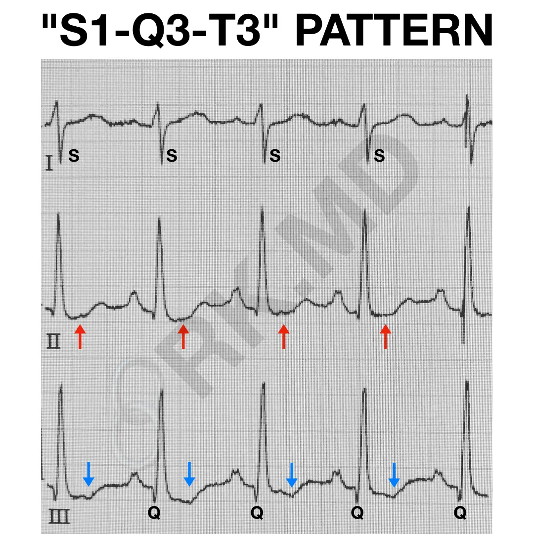
S1Q3T3 EKG Pattern RK.MD
pulmonary disease pattern ecg Hình ảnh có liên quan Diseases Club
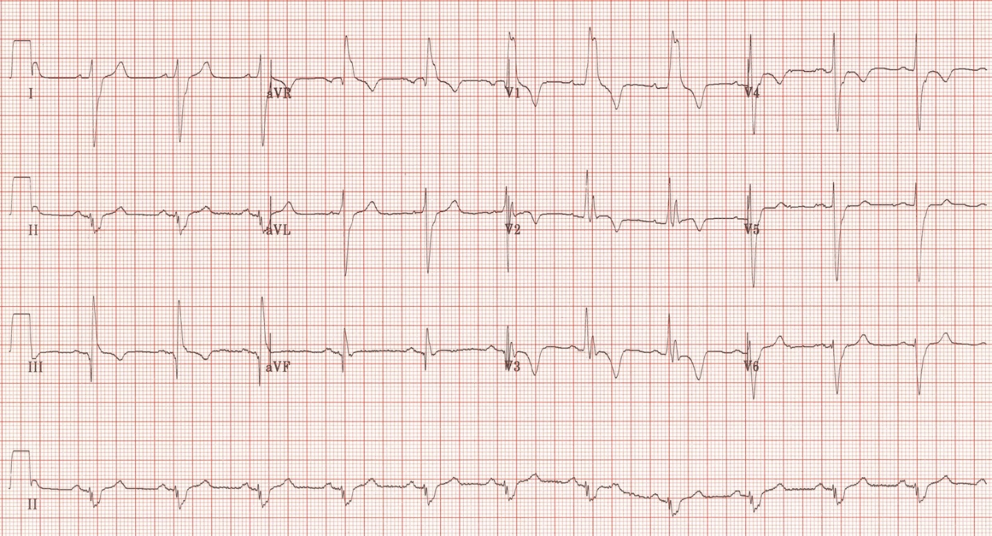
ECG in Chronic Obstructive Pulmonary Disease • LITFL • ECG Library
ECG in Chronic Obstructive Pulmonary Disease • LITFL • ECG Library

The ECG's of Pulmonary Embolism Resus

ECG in Chronic Obstructive Pulmonary Disease • LITFL • ECG Library

Pulmonary Embolism (PE) Causes, symptoms, diagnosis, treatment
Pulmonary Embolism ECG New Health Advisor

Pulmonary Disease Pattern On Ekg Pattern.rjuuc.edu.np

S1Q3T3 on ECG in a patient with Acute Pulmonary Embolism GrepMed
(See Also Electrocardiography In Cardiovascular Disorders.)
•Right Axis Deviation Or Vertical Axis Of The Qrs Complex.
Ecg Findings Often Suggest Right Ventricular Pressure Overload Or Strain.
The Presence Of Hyperexpanded Emphysematous Lungs Within The Chest;
Related Post: