Sine Wave Pattern Ecg
Sine Wave Pattern Ecg - Web hyperkalemia with sine wave pattern. Figure 1 (below) shows normal sinus rhythm at paper speed 25 mm/s. Web the progressively widened qrs eventually merges with the t wave, forming a sine wave pattern. Ecg changes generally do not manifest until there is a moderate degree of hyperkalaemia (≥ 6.0 mmol/l). Web this is the “sine wave” rhythm of extreme hyperkalemia. This is certainly alarming because sine wave pattern usually precedes ventricular fibrillation. Web there are three ecg patterns associated with brugada syndrome, of which only the type 1 ecg is diagnostic. Web the ecg changes reflecting this usually follow a progressive pattern of symmetrical t wave peaking, pr interval prolongation, reduced p wave amplitude, qrs complex widening, sine wave formation, fine ventricular fibrillation and asystole. Web ecg in emergency medicine and acute care 1e, 2004. Web in these situations, the p wave is regular with a constant morphology, but there is either a recurring pattern to the pr interval with intermittent dropped beats (second degree av block) or no relationship at all between p waves and qrs complexes (third degree av block). There is frequently a background progressive bradycardia. Figure 1 (below) shows normal sinus rhythm at paper speed 25 mm/s. Web there are three ecg patterns associated with brugada syndrome, of which only the type 1 ecg is diagnostic. The combination of broadening qrs complexes and tall t waves produces a sine wave pattern on the ecg readout. Tall tented t waves (early sign) prolonged pr interval; Web the ecg changes reflecting this usually follow a progressive pattern of symmetrical t wave peaking, pr interval prolongation, reduced p wave amplitude, qrs complex widening, sine wave formation, fine ventricular fibrillation and asystole. An ecg is an essential investigation in the context of hyperkalaemia. Web several factors may predispose to and promote potassium serum level increase leading to typical electrocardiographic abnormalities. The earliest manifestation of hyperkalaemia is an increase in t wave amplitude. Definition (criteria) for sinus rhythm. Cardiovascular collapse and death are imminent. Web in these situations, the p wave is regular with a constant morphology, but there is either a recurring pattern to the pr interval with intermittent dropped beats (second degree av block) or no relationship at all between p waves and qrs complexes (third degree av block). Sine wave pattern (late sign) arrhythmias The. This pattern usually appears when the serum potassium levels are well over 8.0 meq/l. Sine wave, ventricular fibrillation, heart block; Regular rhythm with ventricular rate between 50 and 100 beats/min. In addition, the t waves are symmetric (upstroke and downstroke equal) (┴), which further supports hyperkalemia as the etiology. Changes not always predictable and sequential. Web the ecg changes reflecting this usually follow a progressive pattern of symmetrical t wave peaking, pr interval prolongation, reduced p wave amplitude, qrs complex widening, sine wave formation, fine ventricular fibrillation and asystole. Web several factors may predispose to and promote potassium serum level increase leading to typical electrocardiographic abnormalities. Web hyperkalaemia is defined as a serum potassium level. Cardiovascular collapse and death are imminent. In addition, the t waves are symmetric (upstroke and downstroke equal) (┴), which further supports hyperkalemia as the etiology. Web the ecg changes reflecting this usually follow a progressive pattern of symmetrical t wave peaking, pr interval prolongation, reduced p wave amplitude, qrs complex widening, sine wave formation, fine ventricular fibrillation and asystole. Web. The morphology of this sinusoidal pattern on ecg results from the fusion of wide qrs complexes with t waves. Web this article deals mainly with ecg features of sinus rhythm. In addition, the t waves are symmetric (upstroke and downstroke equal) (┴), which further supports hyperkalemia as the etiology. This pattern usually appears when the serum potassium levels are well. The morphology of this sinusoidal pattern on ecg results from the fusion of wide qrs complexes with t waves. Web a very wide qrs complex (up to 0.22 sec) may be seen with a severe dilated cardiomyopathy and this is a result of diffuse fibrosis and slowing of impulse conduction. Web sine wave pattern in hyperkalemia is attributed to widening. High serum potassium can lead to alterations in the waveforms of the surface electrocardiogram (ecg). Web ecg in emergency medicine and acute care 1e, 2004. There is frequently a background progressive bradycardia. Web the ecg changes reflecting this usually follow a progressive pattern of symmetrical t wave peaking, pr interval prolongation, reduced p wave amplitude, qrs complex widening, sine wave. Subsequent ventricular fibrillation (vf) or asystole may then follow. Web the ecg changes reflecting this usually follow a progressive pattern of symmetrical t wave peaking, pr interval prolongation, reduced p wave amplitude, qrs complex widening, sine wave formation, fine ventricular fibrillation and asystole. Web the progressively widened qrs eventually merges with the t wave, forming a sine wave pattern. In. Web sine wave pattern in hyperkalemia is attributed to widening of qrs with st elevation and tented t wave merging together with loss of p wave and prolongation of pr interval (ettinger et al., 1974). The earliest manifestation of hyperkalaemia is an increase in t wave amplitude. Web hyperkalemia with sine wave pattern. We describe the case of a patient. Based on lab testing (>5.5 meq/l), although ecg may provide earlier information The morphology of this sinusoidal pattern on ecg results from the fusion of wide qrs complexes with t waves. Peaked t waves, prolonged pr interval, shortened qt interval; The physical examination was unremarkable, but oxygen saturation was. Web in these situations, the p wave is regular with a. Tall tented t waves (early sign) prolonged pr interval; Web a very wide qrs complex (up to 0.22 sec) may be seen with a severe dilated cardiomyopathy and this is a result of diffuse fibrosis and slowing of impulse conduction. We describe the case of a patient who presented with hyperkalaemia and an electrocardiographic aspect consistent with a. Ecg changes generally do not manifest until there is a moderate degree of hyperkalaemia (≥ 6.0 mmol/l). There is frequently a background progressive bradycardia. The morphology of this sinusoidal pattern on ecg results from the fusion of wide qrs complexes with t waves. Regular rhythm with ventricular rate between 50 and 100 beats/min. Web ecg in emergency medicine and acute care 1e, 2004. Sine wave pattern (late sign) arrhythmias Free intro classexpert instructionall levels of expertiseeasy to understand Web several factors may predispose to and promote potassium serum level increase leading to typical electrocardiographic abnormalities. As k + levels rise further, the situation is becoming critical. The earliest manifestation of hyperkalaemia is an increase in t wave amplitude. This is certainly alarming because sine wave pattern usually precedes ventricular fibrillation. An ecg is an essential investigation in the context of hyperkalaemia. Web ecg changes in hyperkalaemia.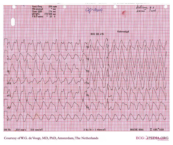
Sine wave pattern wikidoc

12 lead EKG showing sinewave done in the emergency room. Download

SineWave Pattern Arrhythmia and Sudden Paralysis That Result From
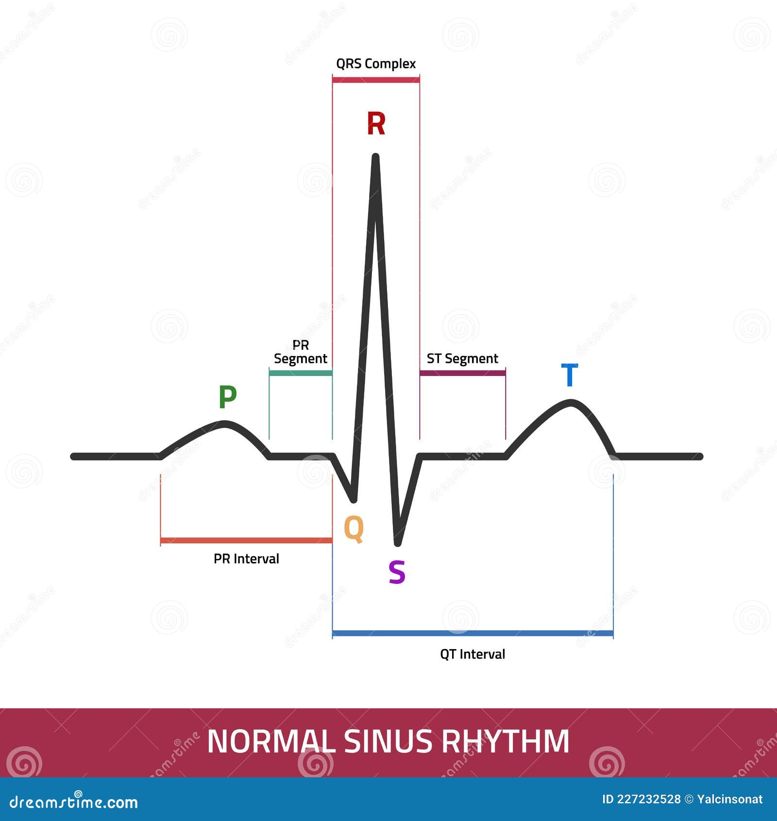
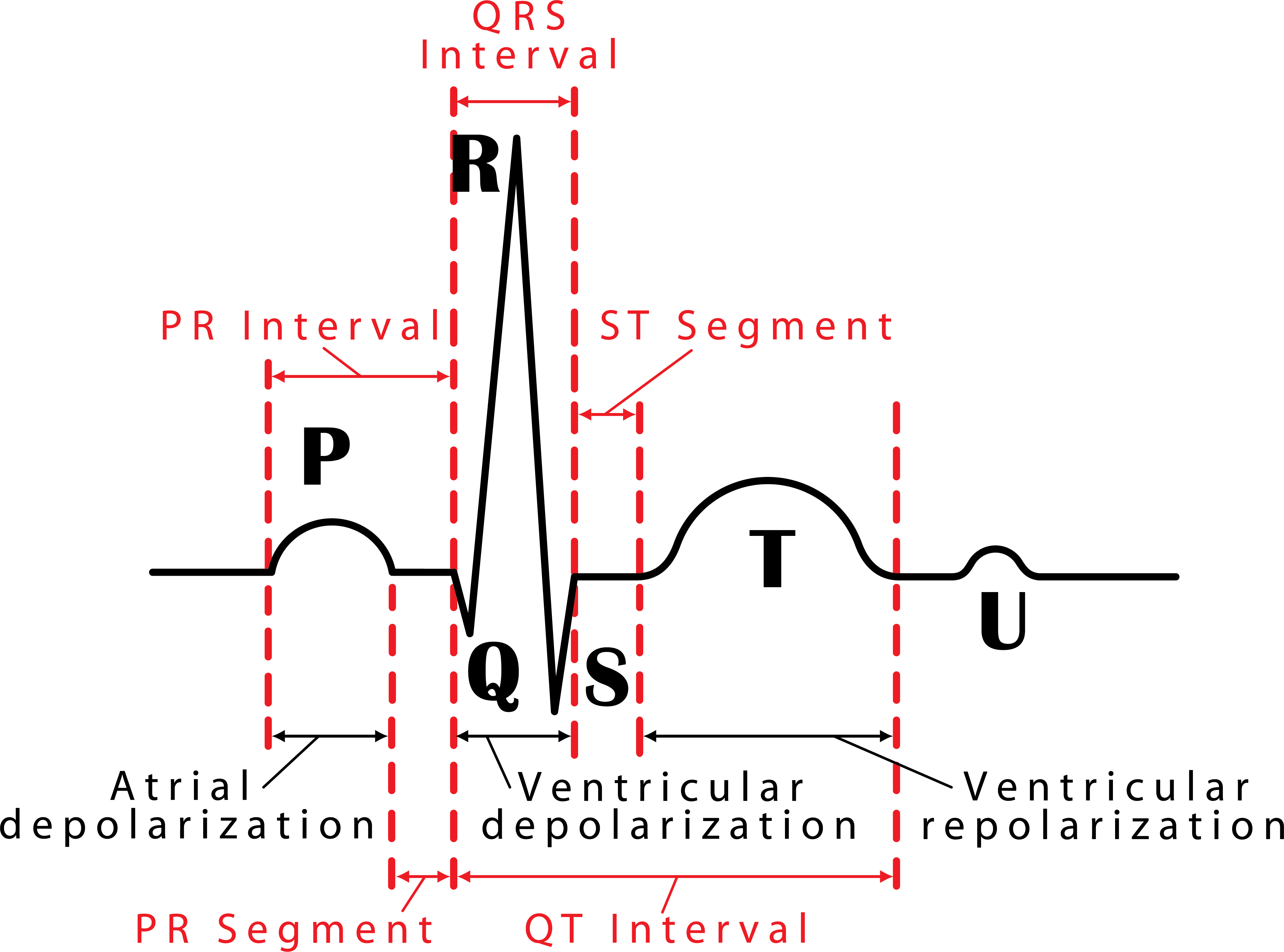
Ecg Normal Sinus Rhythm Infographic Diagram Stock Illustration Images

Hyperkalemia; Hyperpotassemia

Dr. Smith's ECG Blog Weakness and Dyspnea with a Sine Wave. It's not
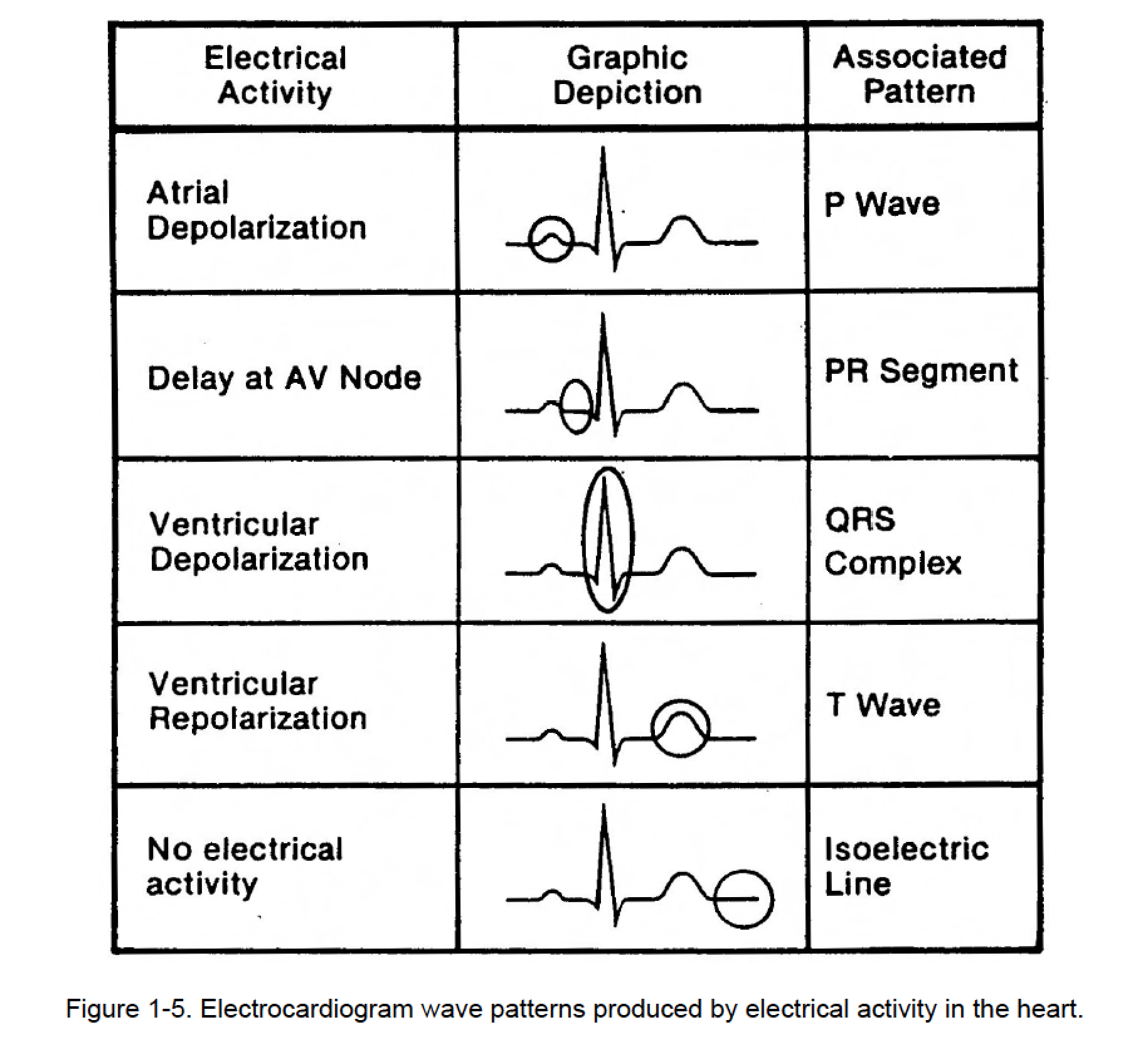
105. GRAPHIC DISPLAY OF ELECTROCARDIOGRAM (C) Cardiac Rhythm

Sine Wave Pattern Ecg Images and Photos finder

ECG changes due to electrolyte imbalance (disorder) Cardiovascular
048 How to Read an Electrocardiogram (ECG/EKG) Interactive Biology
Changes Not Always Predictable And Sequential.
Web This Is The “Sine Wave” Rhythm Of Extreme Hyperkalemia.
Web The Ecg Changes Reflecting This Usually Follow A Progressive Pattern Of Symmetrical T Wave Peaking, Pr Interval Prolongation, Reduced P Wave Amplitude, Qrs Complex Widening, Sine Wave Formation, Fine Ventricular Fibrillation And Asystole.
Web Hyperkalaemia Is Defined As A Serum Potassium Level Of > 5.2 Mmol/L.
Related Post: